Institute of Science, NIRMA University, Ahmedabad.
Background
A miscarriage or spontaneous abortion is the natural death of an embryo or fetus in the womb. Occurrence of miscarriage ranges between 15-30% of all known pregnancies (Stovall et al., 2002).Studies performed on abortuses have consistently shown that approximately 50% are associated with fetal chromosome abnormalities. The sex chromosomal abnormalities arises due to maternal/ paternal meiotic errors, The maternal versus paternal meiotic error vary substantially among the different chromosomes with nearly 50% of XXY pregnancies being the result of paternal non-disjunction whereas less than 10% of other trisomies arise by paternal meiotic error. Monosomy X, another large category (~7% of spontaneous abortions), is generally caused by loss of the paternal sex chromosome (Hassold T et al., 1992), which is consistent with the lack of maternal age effect on the incidence of 45X.
ObjectiveStudy of chromosomal abnormalities due to meiotic errors in recurrent spontaneous abortions.
MethodsChromosomal study is done by tissue culture technique, to rule out the abnormality. In cases where no chromosome preparation was possible, multiplex Interphase FISH probe set was used.
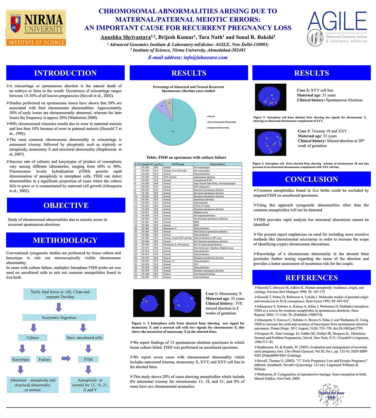
ResultsWe report findings of 48 spontaneous abortion cases (POC samples including fetal and placental tissue). Tissue culture was performed on all 48 specimens. Successful culture and Karyotype was obtained in 14 out of 48 cases, in the remaining cases FISH was performed. We report six cases with chromosomal abnormality which includes autosomal trisomy, monosomy X, XYY, and XXY cell line in the aborted fetus.
ConclusionThis study shows 6.25% of cases showing autosomal trisomy for chromosome 13, 18, and 21; and 8.33% of cases have sex chromosomal anomalies. Identification of genetic cause can have implications for proper genetic counselling and future prenatal diagnosis. Karyotype can unravel hitherto unknown microscopic anomalies; whereas FISH can unravel sub-microscopic anomalies in non-dividing cells also, but requires prior knowledge of possible chromosomal region and is also limited in terms of number of target regions. The present report emphasizes on need for including more sensitive methods like chromosomal microarray in order to increase the scope of identifying cryptic genetic aberrations.